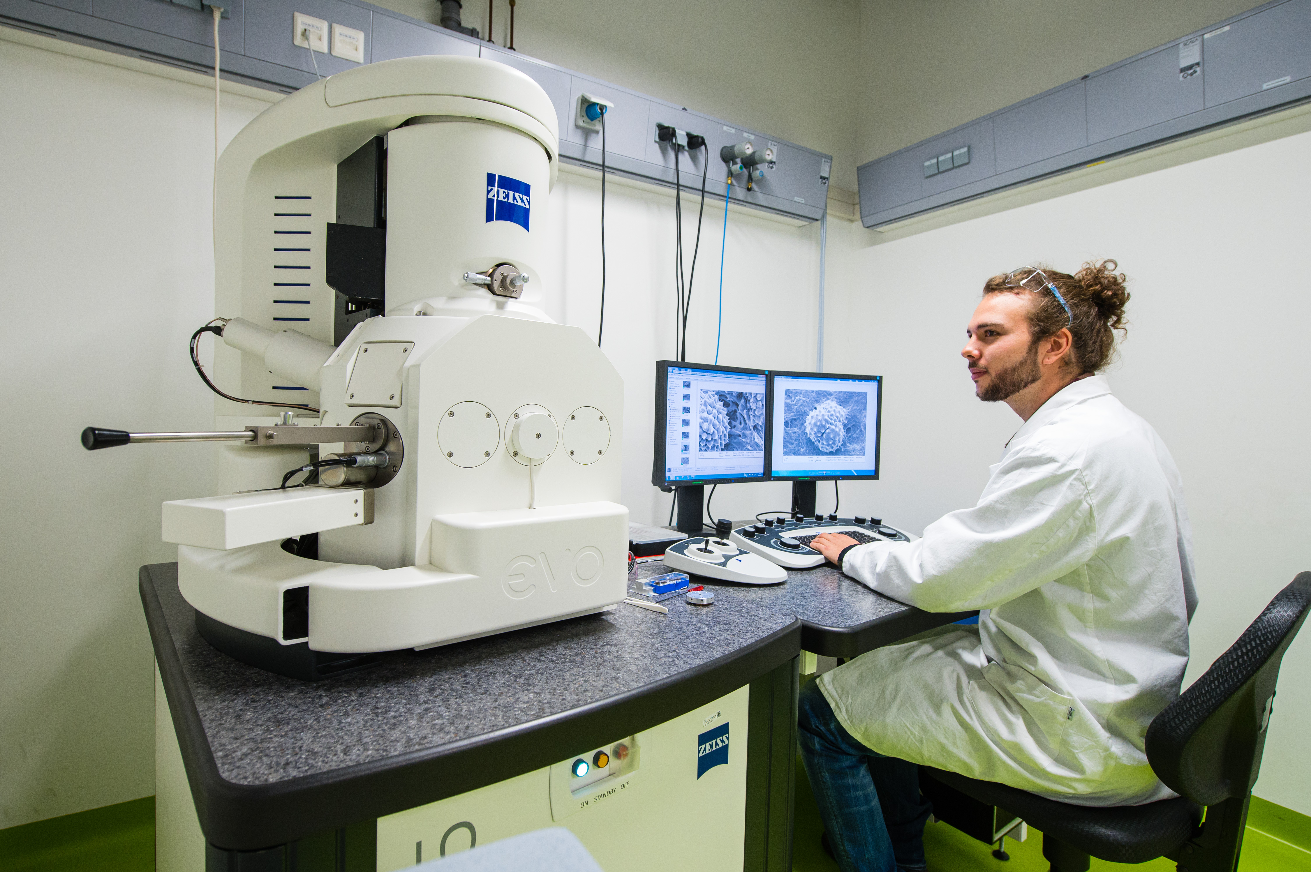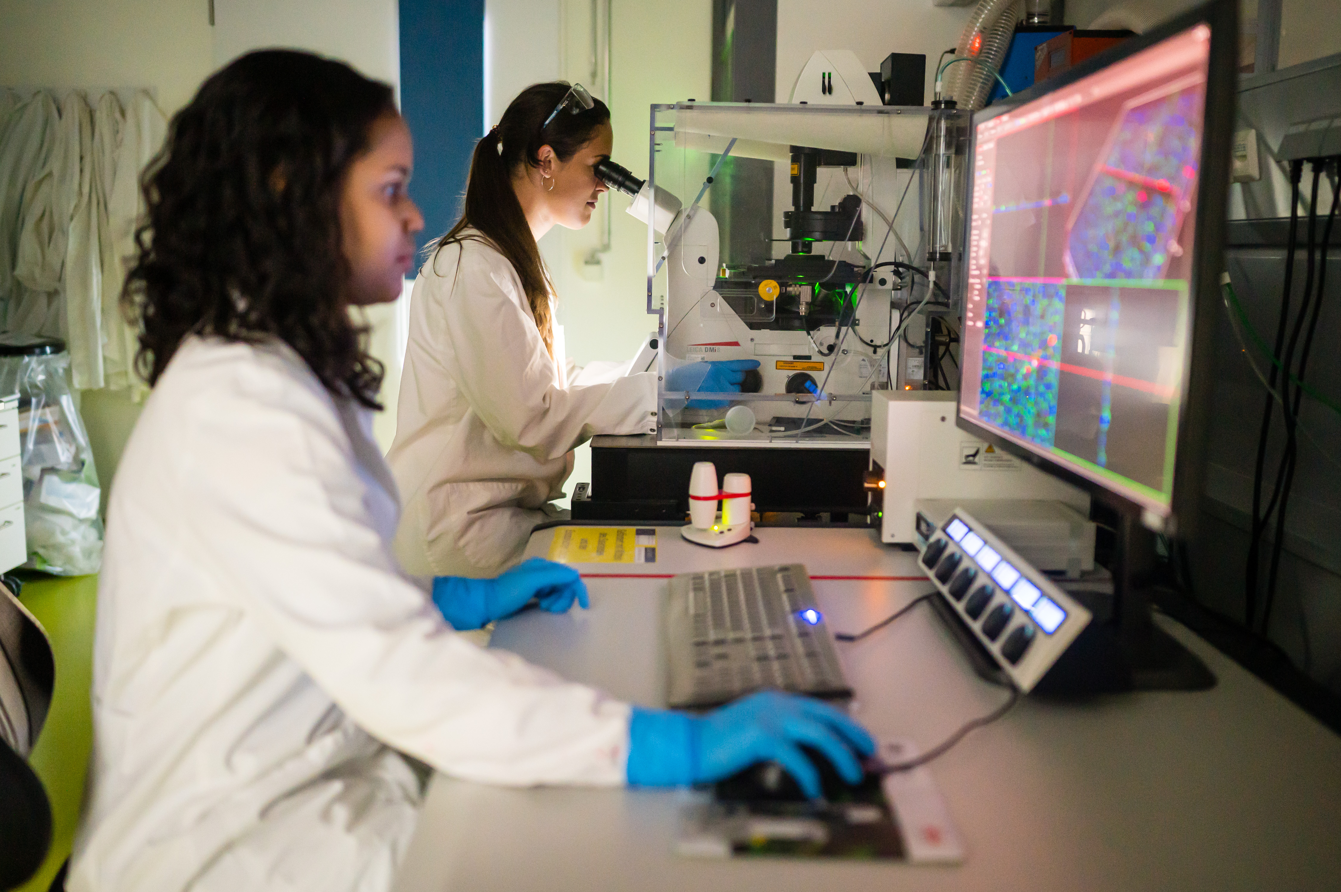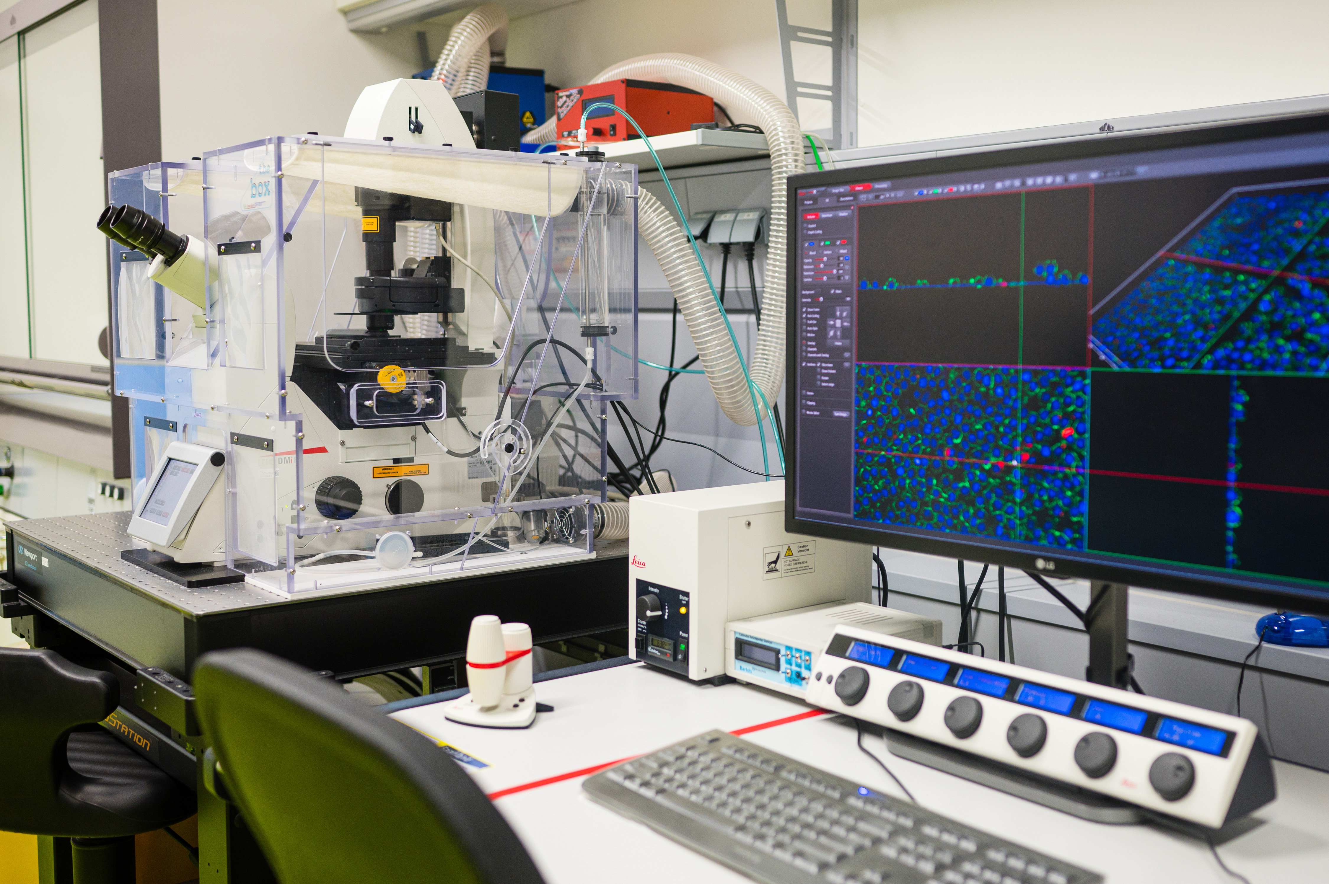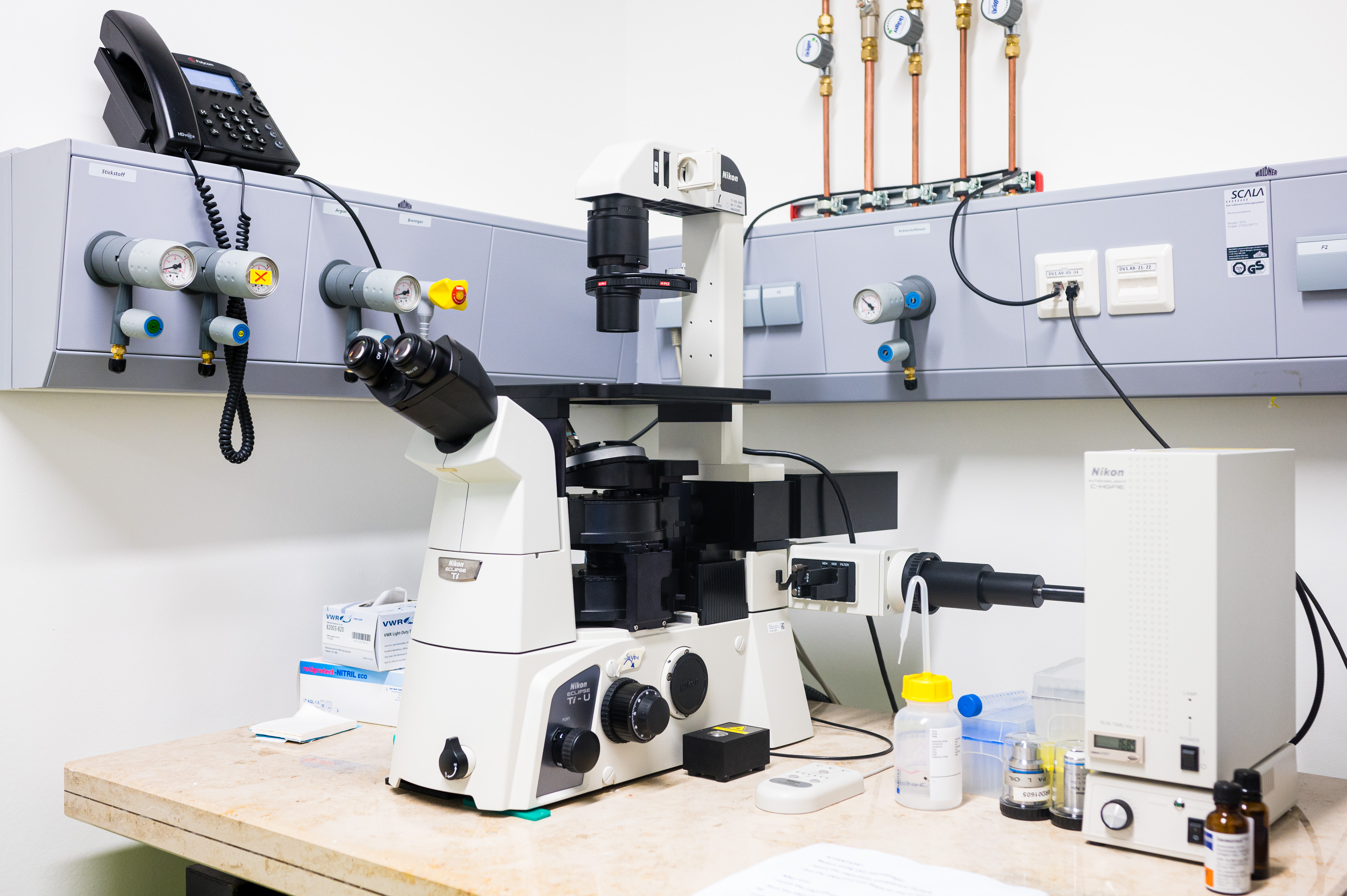
Imaging
The Imaging platform available at HIPS offers cutting-edge technology for both functional and molecular imaging. Combining fluorescence (normal and confocal) and scanning electron microscopes, this platform allows visualising and characterising micro and nanoscale drug delivery systems, as wells as physiological, biochemical and infective phenomena in in vivo/in vitro systems (e.g. living or fixed cells, tissues, biofilms, etc.).
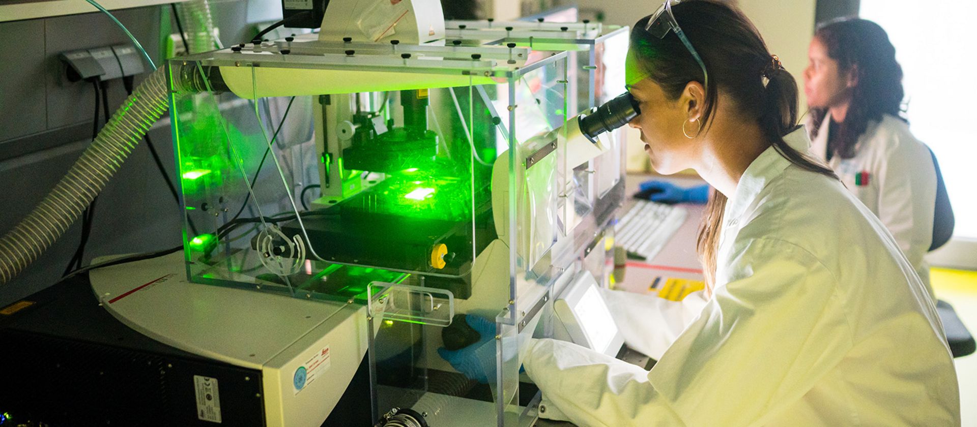
The fluorescence microscope (Nikon Eclipse Ti- S) is an optical microscope that uses fluorescence to observe both biological and inorganic samples. The confocal microscope (Leica DMi8 inverted microscope) also uses fluorescence optics to resolve detailed structures but in several optical planes, bringing the opportunity to obtain high-resolution 3D reconstructions of the samples. Labelling the samples with fluorescent dyes, the fluorescence microscopes allow us to visualise e.g. cellular compartments, cellular growth patterns, biofilm structures or the biodistribution, internalization and subcellular location of drugs and carriers inside the cells or within the tissue.
The scanning electron microscope (SEM EVO HD15) produces high-resolution images and gives information of the sample topography (e.g. shape and size of particles) and composition (e.g. distribution of the nanoparticle size).